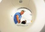
Heart scan software could help save lives
 Biomedical Engineering Biomedical Engineering
New software developed at the University of Adelaide could help to detect and measure cardiac defects in millions of patients right around the world. The software, named MedFlovan, has been developed in collaboration with the University's Centre for Biomedical Engineering, the Cardiovascular Research Centre, and the Cardiovascular Investigation Unit. The developer of this program is PhD student Kelvin Wong from the School of Electrical & Electronic Engineering. Mr Wong has drawn on a range of expertise - including computer science, medicine, physics and mechanical engineering - to develop a new way of quantifying abnormalities in the heart and arteries. "The software is a diagnostic system for detecting cardiac abnormalities and assessing their degree of defects," Mr Wong said. MedFlovan analyses images taken from MRI (magnetic resonance imaging) scans. It measures the changes from one scan to the next and provides vital information about blood flows and turbulence in the heart and arteries. Understanding blood flow patterns in the heart helps to determine the cause of the problem. "This research can be used for identification or quantification of septal defects (a form of congenital heart defect that causes irregular blood flow between the left and right sides of the heart), atherosclerotic arteries (an inflammation or 'hardening' of the arteries), defective heart valves, and certain other cardiac phenomena in the chambers of the heart, such as shunts between the pulmonary and aortic circulations and obstructions inside the heart. "The information can then be used by doctors to diagnose the cardiac problem, determine its severity, and then help to prepare their strategies for the treatment of patients," Mr Wong said. MedFlovan is the culmination of years of hard work by Mr Wong, and a specific desire to do something that benefits society. His presentation on blood flow assessment, based on the software, recently earned him a Young Investigator Award in Singapore at the 15th International Conference on Mechanics in Medicine and Biology. Mr Wong's academic supervisors are Adjunct Professor Jagannath Mazumdar (School of Mathematical Sciences and School of Electrical & Electronic Engineering), Professor Derek Abbott (School of Electrical & Electronic Engineering), Professor Stephen Worthley and Professor Prash Sanders (School of Medicine), and Associate Professor Richard Kelso (School of Mechanical Engineering). His work has also benefited from collaboration with Mr Pawel Kuklik, who is currently a Research Fellow at the Royal Adelaide Hospital. "There are many aspects of MRI technology that are unexplored," Mr Wong said. "It takes a lot of dedication and motivation to try to understand MRI in depth. We are fortunate to have had advice and expertise from various specialists in this area, from the field of computer vision, medical imaging and engineering perspectives. "Various systems have previously been developed for analysing blood flow in arteries and the aorta - they are more specifically designed to do real-time measurement and to physically measure the properties, and not computationally measure it. These other systems are successful and are widely researched, so not many people have focused on the feasibility of developing the type of solution that we proposed," he said. There has been some interest in this work from MRI manufacturers and medical centres, which bodes well for the future commercialisation of the software. To help organise the protection of this research and prepare it for commercialisation, Adelaide Research & Innovation (ARI), the University of Adelaide's commercial development company, has been assisting the research team and has recently filed a provisional patent. Story by David Ellis
|


