Case study: A better life with cystic fibrosis, one breath at a time

Have you ever held your breath for longer than what feels comfortable? Could you feel the overwhelming urge to breathe? This is what people living with cystic fibrosis can feel. As the disease progresses, the feeling gets worse, breath by breath.
The Cystic Fibrosis Airway Research Group at the Robinson Research Institute, led by Associate Professor David Parsons and Associate Professor Martin Donnelley, has made it their mission to keep the genetically inherited, life-threatening disease at bay by developing a novel lung imaging technology that paves the way for early detection and precisely targeted treatment.
“With conditions like cystic fibrosis, early detection of changes in lung function means that a lifetime of complications may be avoided.”Associate Professor David Parsons, co-leader of the Cystic Fibrosis Airway Research group
To treat children with cystic fibrosis, it is critical to know the location and extent of abnormal airflow in the lungs. However, this is the present knowledge gap, particularly when it comes to young children. Current lung function tests such as spirometry, or imaging methods like computed tomography (CT), come with big limitations. Spirometry generally cannot be performed reliably by young children under the age of six. CT scans deliver a high radiation dose and the images lack the precision required, meaning the beginning of disease in young children often goes undetected, robbing them of a chance at preventative treatments.
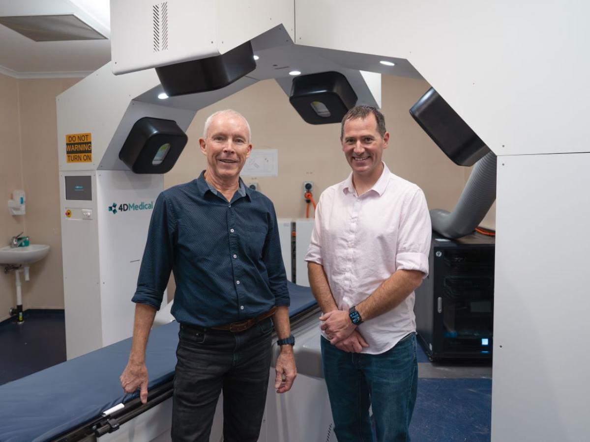
From left: Associate Professor David Parsons, Associate Professor Martin Donnelley.
Closing the gap
X-ray velocimetry (XV) is set to close the gap. This novel imaging technology has been developed to deliver high precision 4D imaging of the lungs. 4D imaging combines three-dimensional (3D) images of the volume of the lungs with the added fourth dimension of time. This enables doctors to visualise where air is flowing inside the lungs – something that was not possible before.
Combining expertise across physics, medicine and engineering, the interdisciplinary team of researchers from the Robinson Research Institute in collaboration with the Women’s and Children’s Hospital in Adelaide, Monash University and industry partner 4DMedical Pty Ltd were awarded the 2023 Aspire Scholarship Eureka Prize for Excellence in Interdisciplinary Scientific Research, for their development and application of XV imaging to cystic fibrosis.
Real world impact
The technology can be considered a game-changer in lung imaging in general and it is particularly promising for young children with cystic fibrosis. Although born with healthy lungs, cystic fibrosis lung disease rapidly causes lung damage in early childhood, initially affecting small patches of the lungs. X-ray velocimetry is capable of detecting the early stages of disease with unparalleled precision, allowing for highly targeted treatment.
“XV lung imaging holds real potential for detecting the earliest beginnings of lung disease in very young children with cystic fibrosis. Since XV can show exactly where in the lung disease is beginning, it means prevention of disease can become a reality, by precisely targeting treatment to just those areas affected, before disease can establish.”Associate Professor Martin Donnelley, co-leader of the Cystic Fibrosis Airway Research group
Clinical trials have commenced at the Women’s and Children’s Hospital in Adelaide with a pilot study recruiting children aged 3–18 years to have x-ray velocimetry lung imaging performed. The study aims to recruit 20 children with healthy lungs and 20 children with cystic fibrosis and is estimated to run until the end of 2024. A second study examining changes in lung function in children taking the new cystic fibrosis drug Trikafta is expected to commence soon and plans to expand trials across a range of paediatric lung diseases are in development.