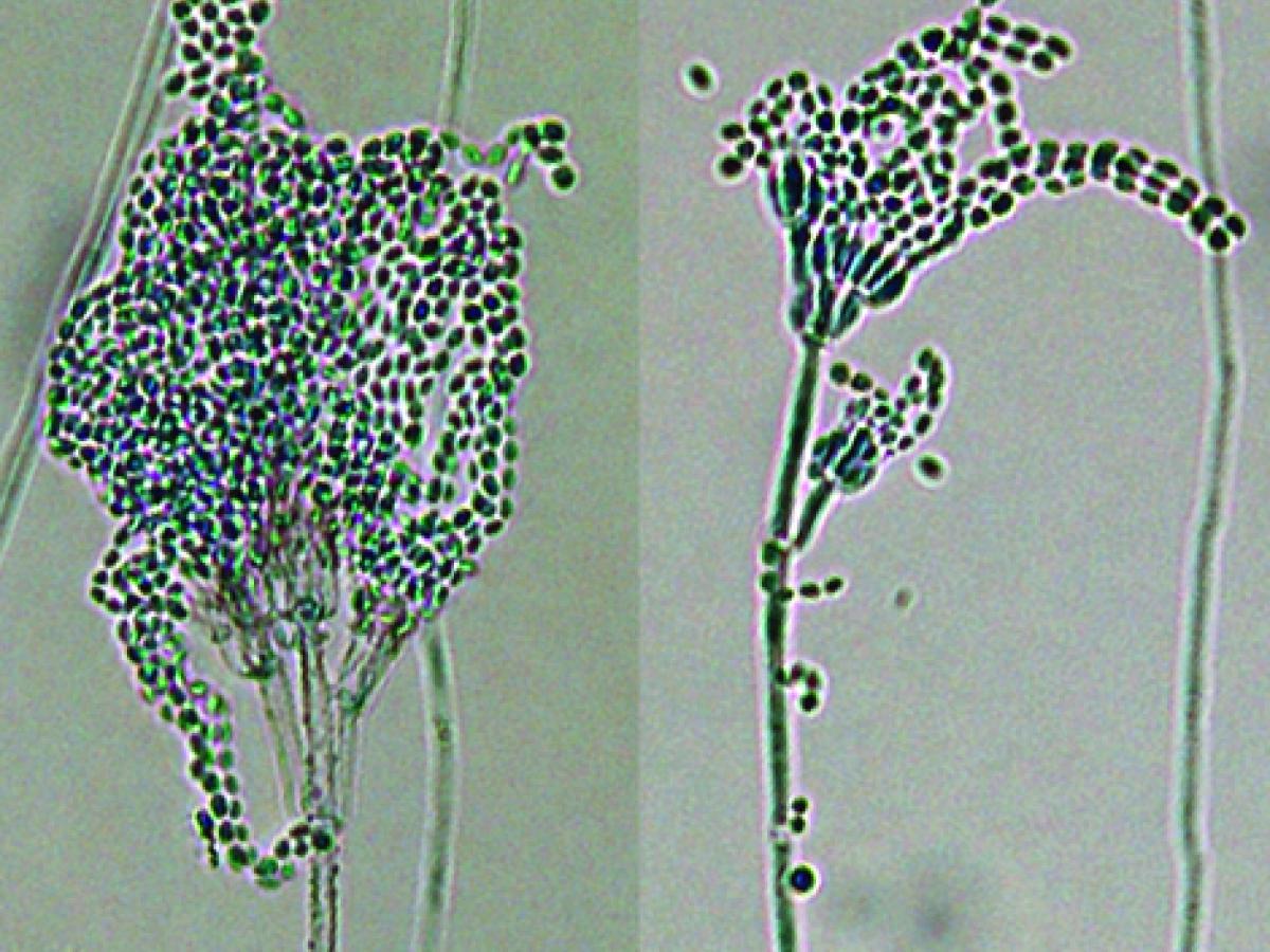
Purpureocillium lilacinum
Synonymy:
Paecilomyces lilacinus
Purpureocillium lilacinum is commonly isolated from soil, decaying vegetation, insects, nematodes and as a laboratory contaminant. It is also a causative agent of infection in human and other vertebrates (Luangsa-ard et al. 2011).
Note: Purpureocillium lilacinum and Marquandomyces marquandii were previously classified within the genus Paecilomyces.
RG-1 organism.

Purpureocillium lilacinum culture.
Morphological description:
Colonies are fast growing, suede-like to floccose, vinaceous to violet-coloured. Conidiophores are erect 400-600 µm in length, bearing branches with densely clustered phialides. Conidiophore stipes are 3-4 µm wide, yellow to purple and rough-walled. Phialides are swollen at their bases, gradually tapering into a slender neck. Conidia are ellipsoidal to fusiform, smooth-walled to slightly roughened, hyaline to purple in mass, 2.5-3.0 x 2-2.2 µm, and are produced in divergent chains. Chlamydospores are absent. Growth at 38C.

Purpureocillium lilacinum conidiophores, phialides and conidia. Note: Rough-walled conidiophore.
Molecular identification:
ITS sequencing is recommended (Atkins et al. 2005, Luangsa-ard et al. 2011).
Key features:
Colony pigmentation, phialides with swollen bases, pigmented and rough-walled conidiophore stipes, absence of chlamydospores and growth at 37C. Note: Paecilomyces marquandii differs by having a yellow reverse pigment, smooth conidiophore stipes, presence of chlamydospores, and no growth at 37C.
References:
Samson (1974), Domsch et al. (1980), McGinnis (1980), Onions et al. (1981), Rippon (1988), de Hoog et al. (2000, 2015), Perdomo et al. (2013).
| Antifungal susceptibility: Purpureocillium lilacinum (Australian national data); MIC µg/mL. | ||||||||||||
|---|---|---|---|---|---|---|---|---|---|---|---|---|
| No | ≤0.03 | 0.06 | 0.125 | 0.25 | 0.5 | 1 | 2 | 4 | 8 | 16 | ≥32 | |
| AmB | 127 | 1 | 1 | 7 | 8 | 110 | ||||||
| ISAV | 26 | 8 | 16 | 2 | ||||||||
| VORI | 125 | 8 | 65 | 43 | 6 | 2 | 1 | |||||
| POSA | 112 | 8 | 15 | 15 | 62 | 11 | 1 | |||||
| ITRA | 128 | 1 | 1 | 12 | 27 | 51 | 8 | 9 | 4 | 13 | ||
