Talaromyces marneffei
Synonymy:
Penicillium marneffei
The genus Talaromyces has been redefined by combining Penicillium subgenus Biverticillium into Talaromyces based upon phylogenetic analysis of the ITS and RPB1 loci (Samson et al. 2011b).
The genus contains 88 species that were placed into seven sections based on a multigene phylogeny of the ITS, β-tubulin and RPB2 regions (Yilmaz et al. 2014). T. marneffei is the only known dimorphic species in the genus, producing filamentous growth at 25C and a yeast phase at 37C (Andrianopoulos 2002).
WARNING: RG-3 organism.
Cultures of Talaromyces marneffei may represent a biohazard to laboratory personnel and should be handled with caution in a class II Biological Safety Cabinet (BSCII). T. marneffei exhibits thermal dimorphism and is endemic in Southeast Asia and the southern region of China.
Molecular identification:
ITS sequencing is recommended, as well as β-tubulin as a secondary molecular marker for identification (Yilmaz et al.2014).
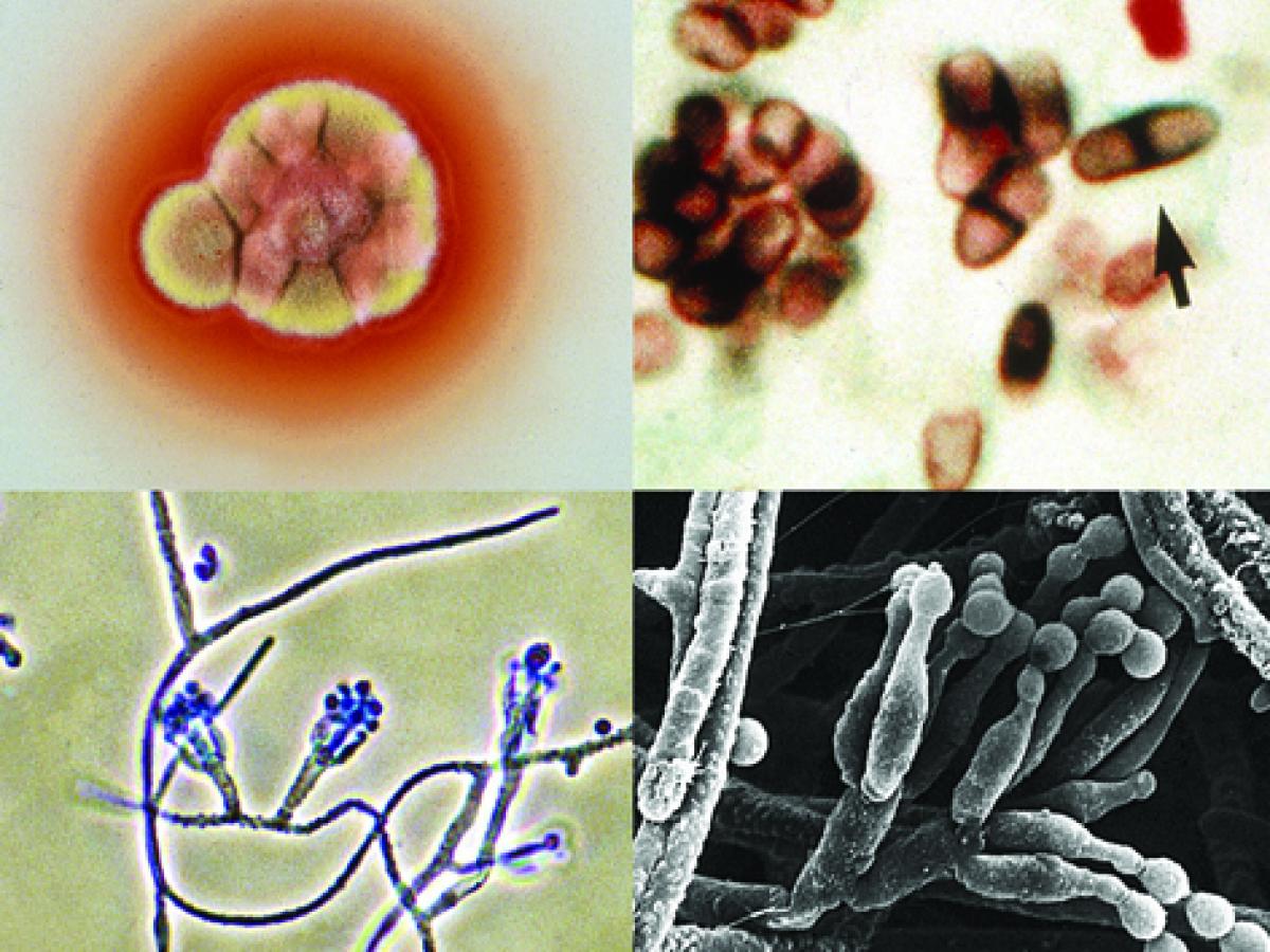
Talaromyces marneffei culture showing red soluble pigment; a giemsa stained touch smear showing typical septate yeast-like cells (arrow), phialides and conidia.
Morphological description:
Colonies at 25C are fast growing, suede-like to downy, white with yellowish-green conidial heads. Colonies become greyish-pink to brown with age and produce a diffusible brownish-red to wine-red pigment. Conidiophores generally biverticillate and sometimes monoverticillate; hyaline, smooth-walled and bear terminal verticils of three to five metulae, each bearing three to seven phialides. Phialides are acerose to flask-shaped. Conidia are globose to subglobose, 2-3 µm in diameter, smooth-walled and are produced in basipetal succession from the phialides.
On brain heart infusion (BHI) agar containing blood incubated at 37C, colonies are rough, glabrous, tan-coloured and yeast-like. Microscopically, yeast cells are spherical to ellipsoidal, 2-6 µm in diameter, and divide by fission rather than budding. Numerous short hyphal elements are also present.
Histopathology:
Tissue sections show small, oval to ellipsoidal yeast-like cells, 3 µm in diameter, either packed within histiocytes or scattered through the tissue. Occasional, large, elongated sausage-shaped cells, up to 8 µm long, with distinctive septa may be present.
Key features:
Talaromyces marneffei is the only dimorphic species of Talaromyces, which grows as a yeast at 37C. It produces a red soluble pigment on general media and conidiophores have flask-shaped to acerose phialides.
References:
Pitt (1979), Ramirez (1982), de Hoog et al. (2000, 2015), Andrianopoulos (2002), Lyratzopoulos et al. (2002), Samson et al. (2011b), Visagie et al. (2014), Yilmaz et al. (2014).
| No | ≤0.016 | 0.03 | 0.06 | 0.125 | 0.25 | 0.5 | 1 | 2 | 4 | ≥8 | |
|---|---|---|---|---|---|---|---|---|---|---|---|
| AmB | 5 | 2 | 2 | 1 | |||||||
| VORI | 5 | 5 | |||||||||
| POSA | 4 | 4 | |||||||||
| ITRA | 5 | 5 |