Mucor
The genus Mucor contains about 50 recognised taxa, many of which have widespread occurrence and are of considerable economic importance (Zycha et al. 1969, Schipper 1978, Domsch et al. 1980).
However, only a few thermotolerant species are of medical importance and human infections are only rarely reported. Most infections reported list M. circinelloides and similar species such as M. indicus, M. ramosissimus, M. irregularis and M. amphibiorum as the causative agents. However, M. hiemalis and M. racemosus have also been reported as infectious agents, although their inability to grow at temperatures above 32C raises doubt as to their validity as human pathogens and their pathogenic role may be limited to cutaneous infections (Scholer et al. 1983, Goodman and Rinaldi 1991, Kwon-Chung and Bennett 1992, de Hoog et al. 2000, 2015).
| Maximum temperature for growth of the reported pathogenic species of Mucor. | ||
|---|---|---|
| Species | Max temp (C) | Pathogenicity |
| M. amphibiorum | 36 | Animals, principally amphibians |
| M. circinelloides | 37 | Animals, occassionally humans |
| M. hiemalis | 30 | Questionable cutaneous infections only |
| M. indicus | 42 | Humans and animals |
| M. irregularis | 38 | Humans |
| M. racemosus | 32 | Questionable |
| M. ramosissimus | 36 | Humans and animals |
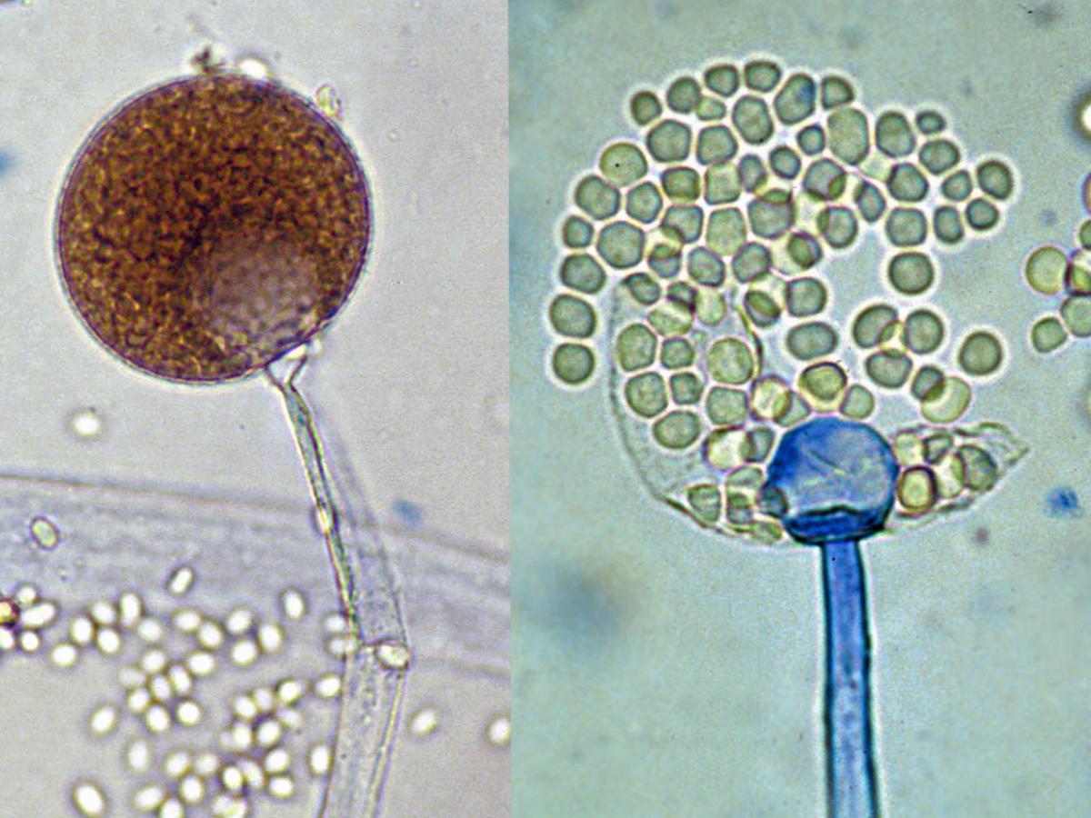
Sporangia, columella and sporangiospores of Mucor spp.
Morphological description:
Colonies are very fast growing, cottony to fluffy, white to yellow, becoming dark-grey, with the development of sporangia. Sporangiophores are erect, simple or branched, forming large (60-300 µm in diameter), terminal, globose to spherical, multispored sporangia, without apophyses and with well-developed subtending columellae. A conspicuous collarette (remnants of the sporangial wall) is usually visible at the base of the columella after sporangiospore dispersal. Sporangiospores are hyaline, grey or brownish, globose to ellipsoidal, and smooth-walled or finely ornamented. Chlamydospores and zygospores may also be present.
Key features:
Mucorales, large, spherical, non-apophysate sporangia with pronounced columellae and conspicuous collarette at the base of the columella following sporangiospore dispersal.
Molecular identification:
ITS sequencing recommended (Walther et al. 2012).
References:
Schipper (1978), Domsch et al. (1980), McGinnis (1980), Onions et al. (1981), Scholer et al. (1983), Rippon (1988), Goodman and Rinaldi (1991), Samson et al. (1995), de Hoog et al. (2000, 2015), Schipper and Stalpers (2003), Ellis (2005b).
Species descriptions
-
Mucor amphibiorum
RG-2 organism.
Morphological description:
Colonies are greyish-brown, slightly aromatic and do not grow at 37C (maximum temperature for growth is 36C). Sporangiophores are hyaline, erect and mostly unbranched, rarely sympodially branched. Sporangia are dark-brown, up to 75 µm in diameter, and are slightly flattened with a diffluent membrane. Columellae are subglobose to ellipsoidal or pyriform, up to 60 x 50 µm, with small collarettes. Sporangiospores are smooth-walled, spherical, and 3.5-5.5 µm in diameter. Zygospores, when formed by compatible mating types, are spherical to slightly compressed, up to 70 x 60 µm in diameter, with stellate projections.Comment:
Mucor amphibiorum is distinguished by poor branching of the sporangiophores and by globose sporangiospores. Ethanol and nitrates are not assimilated (Schipper 1978, Scholer et al. 1983, Hoog et al.2000, 2015).Antifungal susceptibility: Mucor amphibiorum very limited data (Australian national data); MIC µg/mL. Antifungal No 0.016 0.03 0.06 0.125 0.25 0.5 1 2 4 ≥8 AmB 2 1 1 VORI 2 2 POSA 2 1 1 ITRA 2 1 1 -
Mucor circinelloides
M. circinelloides is a common and variable species that includes four formae: circinelloides, lusitanicus, griseocyanus and janssenii (Schipper 1978, Scholer et al. 1983).
RG-1 organism.
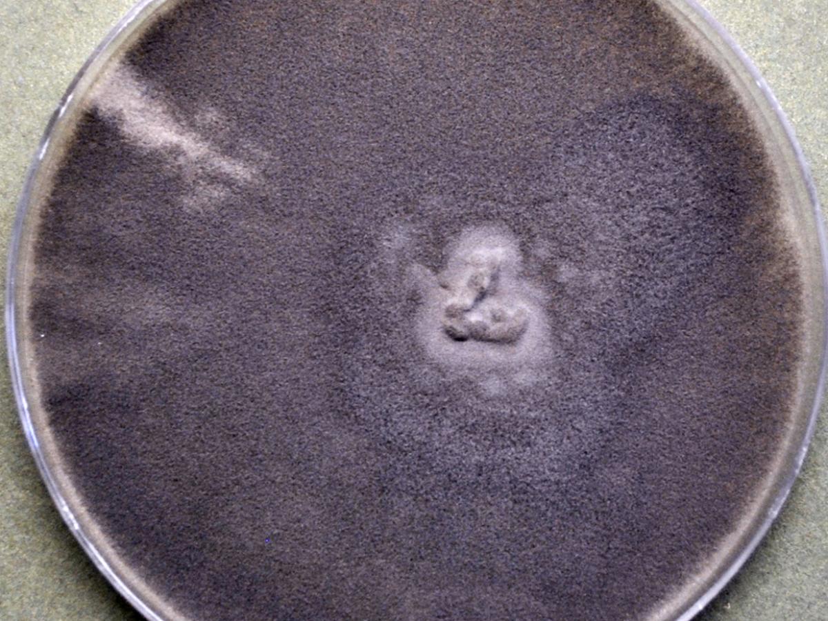
Mucor circinelloides culture.
Morphological description:
Colonies are floccose, pale greyish-brown and grow poorly at 37C (maximum growth temperature 37C). Sporangiophores are hyaline and mostly sympodially branched with long branches erect and shorter branches becoming circinate (coiled). Sporangia are spherical, varying from 20-80 µm in diameter, with small sporangia often having a persistent sporangial wall. Columellae are spherical to ellipsoidal and are up to 50 µm in diameter. Sporangiospores are hyaline, smooth-walled, ellipsoidal, and 4.5-7 x 3.5-5 µm in size. Chlamydospores are generally absent. Zygospores are only produced in crosses of compatible mating types and are reddish-brown to dark-brown, spherical with stellate spines, up to 100 µm in diameter and have equal to slightly unequal suspensor cells.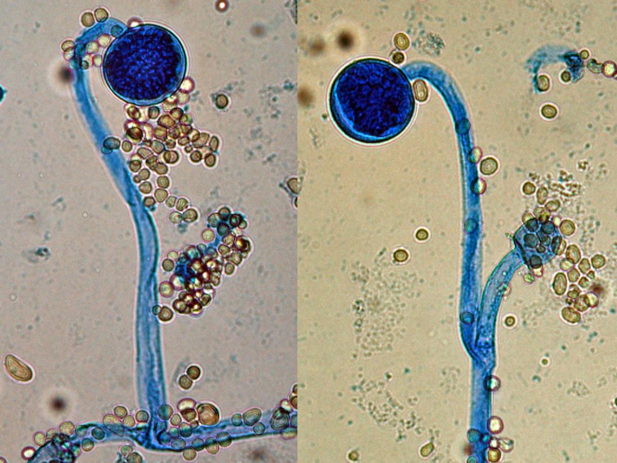
Mucor circinelloides sporangia showing circinate sporangiophores, also note the columella with inconspicuous collarette and sporangiospores.
Comment:
M. circinelloides differs from other species of Mucor in its formation of short circinated, branched sporangiophores bearing brown sporangia and its ability to assimilate ethanol and nitrates (Schipper 1976, Scholer et al. 1983, Samson et al. 1995, de Hoog et al. 2000, Schipper and Stalpers 2003).Antifungal susceptibility: Mucor circinelloides very limited data (Espinel-Ingroff et al., 2015a; Pfaller et al., 2018 and Australian national data); MIC µg/mL. Antifungal No 0.016 0.03 0.06 0.125 0.25 0.5 1 2 4 8 16 ≥32 AmB 154 1 4 17 54 58 20 ISAV 67 1 10 22 25 9 VORI 30 30 POSA 151 2 2 10 30 63 31 5 4 4 ITRA 80 4 6 26 16 15 5 7 -
Mucor indicus
RG-1 organism.
Morphological description:
Colonies are characteristically deep-yellow, aromatic and have a maximum growth temperature of 42C. Sporangiophores are hyaline to yellowish, erect or rarely circinate and repeatedly sympodially branched, with long branches. Sporangia are yellow to brown, up to 75 µm in diameter, with diffluent membranes. Columellae are subglobose to pyriform, often with truncate bases, up to 40 µm high. Sporangiospores are smooth-walled, subglobose to ellipsoidal, and 4-5 µm in diameter. Chlamydospores are produced in abundance, especially in the light. Zygospores are black, spherical up to 100 µm in diameter, with stellate spines and unequal suspensor cells.Comment:
Mucor indicus differs from other species of Mucor by its characteristic deep-yellow colony colour, growth at over 40C, assimilating ethanol, but not nitrate, and thiamine dependence (Schipper 1978, de Hoog et al. 2000, Schipper and Stalpers 2003).Antifungal susceptibility: Mucor indicus limited data (Espinel-Ingroff et al., 2015a and Australian national data); MIC µg/mL. Antifungal No 0.016 0.03 0.06 0.125 0.25 0.5 1 2 4 8 16 ≥32 AmB 47 2 7 22 14 2 ISAV 12 1 3 4 4 VORI 39 35 4 POSA 47 3 12 20 10 2 ITRA 37 1 5 19 6 6 -
Mucor irregularis
Synonymy:
Rhizomucor variabilisRG-2 organism.
Morphological description:
Colonies are whitish to ochraceous, with buff-ochre reverse. Sporangiophores arising from hyphae or from stolons; rhizoids abundant. Sporangiophores are hyaline, up to 2 mm long, 9-23 μm wide, simple or once branched, with branches terminating at a higher level than the main stems; branches all ending in a sporangium. Sporangia subspherical to spherical, up to 100 μm diameter. Columella spherical, ellipsoidal to cylindrical, about 40 μm wide, sometimes lobed, with or without an apophysis. Sporangiospores are hyaline, smooth-walled, very variable, mostly subspherical to ellipsoidal, 3-11 × 2-7 μm. Chlamydospores are abundant. Maximum growth temperature 38C.Comment:
Mucor irregularis differs from other species of Mucor by having abundant rhizoids of different sizes and sporangiospores of highly variable shape, mostly subspherical to ellipsoidal (Lu et al. 2009, 2013). This species is closely related to Mucor hiemalis (Voigt et al. 1999). -
Mucor ramosissimus
RG-1 organism.
Morphological description:
Colonial growth is restricted, greyish and does not grow at 37C (maximum temperature for growth is 36C). Sporangiophores are hyaline, slightly roughened, tapering towards the apex and are erect with repeated sympodial branching. Sporangia are grey to black, globose or somewhat flattened, up to 80 µm in diameter and have very persistent sporangial walls. Columellae are applanate (flattened), up to 40-50 µm in size and are often absent in smaller sporangia. Sporangiospores are faintly brown, smooth-walled, subglobose to broadly ellipsoidal, 5-8 x 4.5-6 µm in size. Oidia may be present in the substrate hyphae, chlamydospores and zygospores are absent. Assimilation of ethanol is negative and that of nitrate is positive.Comment:
Mucor ramosissimus differs from other species of Mucor by its low, restricted growth on any medium, extremely persistent sporangial walls, columellae that are applanate or absent in smaller sporangia (often resembling Mortierella species), short sporangiophores that repeatedly branch sympodially as many as 12 times, and the occurrence of racket-shaped enlargements in the sporangiophores (Hesseltine and Ellis 1964b, Schipper 1976, Scholer et al. 1983, de Hoog et al. 2000, Schipper and Stalpers 2003).Antifungal susceptibility: Mucor ramosissimus very limited data (Espinel-Ingroff et al., 2015a and Australian national data); MIC µg/mL. Antifungal No 0.016 0.03 0.06 0.125 0.25 0.5 1 2 4 8 ≥16 AmB 21 2 6 4 7 1 1 VORI 2 2 POSA 2 1 1 ITRA 4 1 2 1