Status message
Correct! Excellent, you have really done well. Please find additional information below.
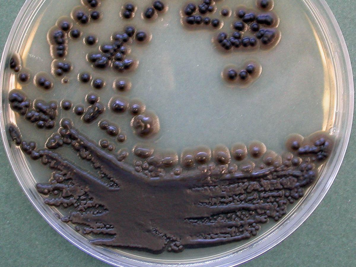
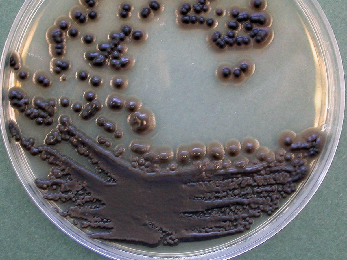
Unknown 18 = Exophiala dermatitidis
Culture: Colonies are slow growing, initially black and yeast-like, becoming suede-like, olivaceous grey and mould-like with age. Cultures grow at 42C.

Microscopy: An initial yeast-like phase is characterized by unicellular, ovoid to elliptical, budding yeast-like cells. With the development of mycelium, flask-shaped to cylindrical annellides are produced. Spherical phialides with large, fragile collarettes may also be present (not seen in this strain). Conidia are hyaline to pale brown, one-celled, round to obovoid, 2.0-4.0 x 2.5-6.0 um, smooth-walled and accumulate in slimy balls (glioconidia) at the apices of the phialides or down their sides.
The identity of this isolate was confirmed by sequencing the the D1/D2 region of large subunit rRNA. The strain showed >99.8% identity to numerous E. dermatitidis strains including strains from CBS while it only shows 97.5% ID to Exophiala jeanselmei.

Comment: Exophiala dermatitidis has been isolated from plant debris and soil and is a recognized causative agent of mycetoma and phaeohyphomycosis in humans. Clinical manifestations include subcutaneous cystic lesions, endocarditis and brain abscesses. E. dermatitidis is neurotropic and cerebral infections are frequently seen.
About Exophiala Back to virtual assessment


