Status message
Correct! Excellent, you have really done well. Please find additional information below.
Unknown 20 = Cladophialophora carrionii
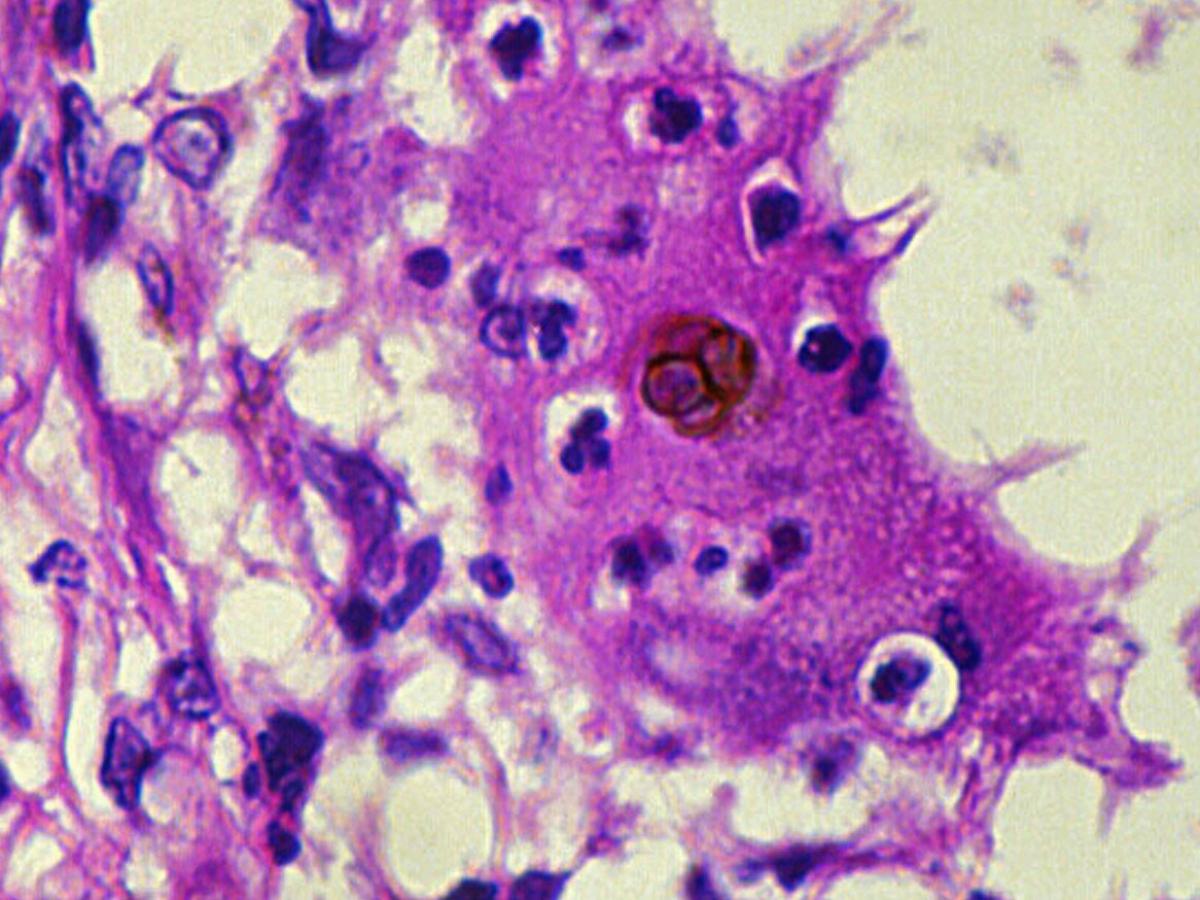
Histopathology: H&E stained section showing characteristic dark brown sclerotic cells which divide by binary fission and not by budding. All agents of chromoblastomycosis form these sclerotic bodies in tissue. Direct microscopy of tissue is necessary to differentiate between chromoblastomycosis and phaeohyphomycosis where the tissue morphology of the causative organism is mycelial.
Culture: On Sabouraud's dextrose agar, colonies are slow growing, reaching 3-4 cm in diameter after one month, with a compact suede-like to downy surface. Colonies are olivaceous-black in colour and have well defined margins.

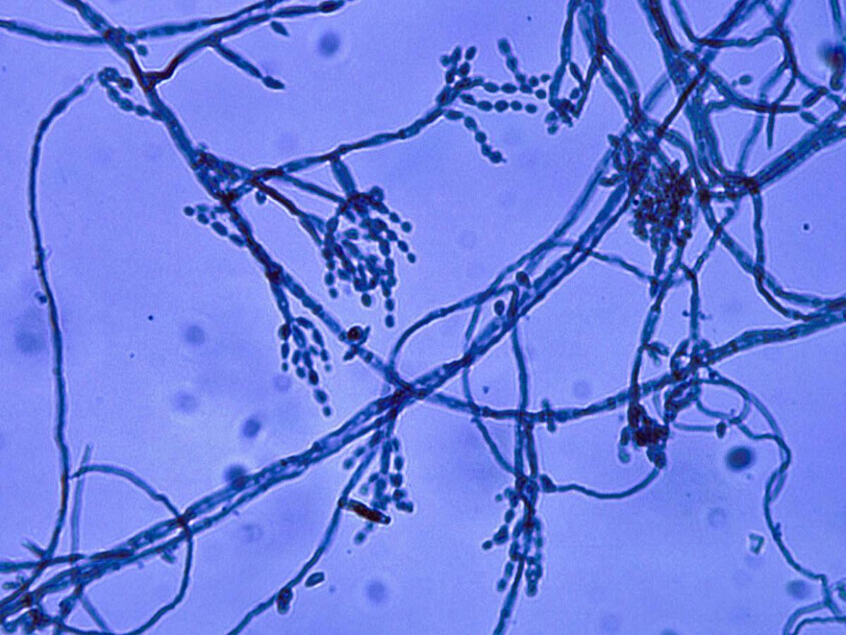
Microscopy: Microscopic morphology shows a Cladosporium type conidial ontogeny with elongate conidiophores producing branched acropetal chains of smooth-walled conidia, 1.5-3.0 x 2.0-7.0 um in size. Maximum growth temperature 35-37C.

Comment: Cladophialophora carrionii is a recognized agent of chromoblastomycosis and it has been isolated from soil and fence posts made from Eucalyptus sp. Cases of chromoblastomycosis caused by C. carrionii are commonly found in Australia, Venezuela, Madagascar and South America. Isolates from phaeomycotic cysts and opportunistic infections have also been reported.
About Cladophialophora Back to virtual assessment