Rhizomucor
The genus Rhizomucor is distinguished from Mucor by the presence of stolons and poorly developed rhizoids at the base of the sporangiophores and by the thermophilic nature of its two species: R. miehei and R. pusillus.
Both of these species are potential human and animal pathogens and were originally classified in the genus Mucor. Rhizomucor pusillus is cosmopolitan and both R. miehei and R. pusillus have been reported as pathogens to humans and animals, the latter to a greater extent.
References:
Cooney and Emerson (1964), Schipper (1978), Domsch et al. (1980), McGinnis (1980), Ellis and Keane (1981), Scholer et al. (1983), de Hoog et al. (2000, 2015), Schipper and Stalpers (2003) and Ellis (2005b).
Identification of most Mucorales is based primarily on the morphology of the sporangia; i.e. arrangement and number of sporangiospores, shape, colour, presence or absence of columellae and apophyses, as well as the arrangement of the sporangiophores and the presence or absence of rhizoids. Growth temperature tests can also be especially helpful in identifying and differentiating members of the genera Rhizomucor, Rhizopus and Lichtheimia.
Species descriptions
-
Rhizomucor miehei
Synonymy: Mucor miehei
This species has been reported as a rare cause of bovine mastitis (Scholer et al. 1983) and is similar in many respects to R. pusillus.
RG-1 organism.
Morphological description:
All strains are homothallic forming numerous zygospores, which are reddish-brown to blackish-brown, globose to slightly compressed, up to 50 µm in diameter, with stellate warts and equal suspensor cells. Colony colour is a dirty grey rather than brown, and sporangia have spiny walls, are up to 50-60 µm in diameter, with columellae rarely larger than 30 µm in diameter. Growth is stimulated by thiamine, with no assimilation of sucrose and maximum growth temperature is 54-58C.Key features:
Growth at 45C, the formation of numerous zygospores, a dirty grey culture colour and a partial growth requirement for thiamine.Antifungal susceptibility: Rhizomucor miehei (Australian national data); MIC µg/mL. Antifungal No 0.016 0.03 0.06 0.125 0.25 0.5 1 2 4 ≥8 AMB 11 3 2 3 2 1 VORI 11 3 5 3 POSA 11 4 4 1 2 ITRA 11 4 5 1 1 -
Rhizomucor pusillus
Synonymy: Mucor pusillus
This species is a rare human pathogen. It has been reported from cases of pulmonary, disseminated and cutaneous types of infection. It is more often associated with animal disease, especially bovine abortion. Rhizomucor pusillus has a worldwide distribution and is commonly associated with compost heaps.
RG-2 organism.
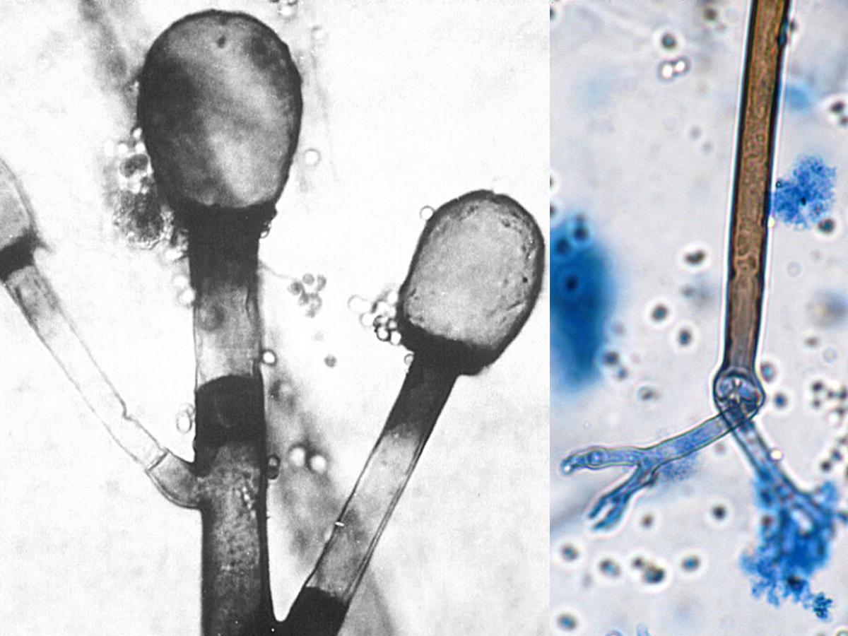
Sporangiophores, columellae and primitive rhizoids of R. pusillus.
Morphological description:
Cultures are characterised by compact, low growing (2-3 mm high), grey to greyish brown-coloured mycelium and by the development of typical sympodially branched, hyaline to yellow-brown sporangiophores (8-15 μm in diameter), always with a septum below the sporangium. Sporangia are globose (40-60 µm in diameter), each possessing an oval or pear-shaped columella (20-30 µm), often with a collarette. Sporangiospores are hyaline, smooth-walled, globose to subglobose, occasionally oval (3-5 µm), and are often mixed with crystalline remnants of the sporangial wall. Chlamydospores are absent. Zygospores are rough-walled, reddish brown to black, 45-65 µm in diameter and may be produced throughout the aerial hyphae in matings between compatible isolates. Temperature growth range: minimum 20-27C; optimum 35-55C; maximum 55C. There is positive assimilation of sucrose and no thiamine dependence.Key features:
Mucorales, growth at 45C (thermophilic), poorly developed stolons and rhizoids, branching sporangiophores with a septum below the sporangium, dark-coloured sporangia without apophyses and smooth-walled globose to subglobose sporangiospores.Antifungal susceptibility: Rhizomucor pusillus ((Espinel-Ingroff et al. 2015a, Australian national data); MIC µg/mL. Antifungal No 0.016 0.03 0.06 0.125 0.25 0.5 1 2 4 ≥8 AMB 37 3 8 9 12 1 2 1 VORI 4 4 POSA 37 1 5 13 10 7 1 ITRA 19 4 5 5 3 1 1